Naber’s probe
Assessment instruments
Furcation Nabers probe and furca classification
According to the glossary of terms of the American Academy of Periodontology, a furcation involvement exists when periodontal disease has caused attachment loss and resorption of bone into the bi- or trifurcation area of a multi-rooted tooth.
Various classification systems have been proposed to describe furcation lesions.
- Classification of the horizontal component of furcation involvement
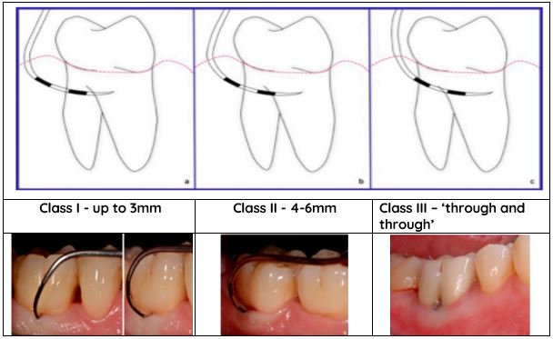
In 1975 Hamp, Nyman and Lindhe proposed a classification system referring to the horizontal attachment loss which is based on three classes:
Class I: horizontal attachment loss < 3 mm of the total width of the furcation area
Class II: horizontal attachment loss > 3 mm but not encompassing the total width of the furcation area
Class III: “through and through” destruction of the periodontal tissue in the furcation area
Naber’s probe is used to determine the extent of the horizontal attachment loss in furcations.
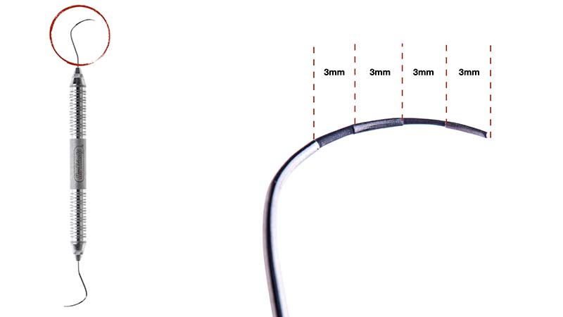
Nabers color-coded probe
Color coded furcation probe
Silver and black increments of 3 mm
Used for probing furcation lesions to evaluate severity of horizontal furcation involvement
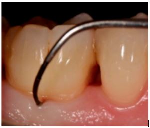
How to assess furcation involvement with Naber’s probe?
Step I
Assessment of possibility of furcation involvement
Possibility of furcation involvement by detecting the probing depth (PD) with the periodontal probe. Clinical attachment loss of 5mm may be indicative of possible furcation involvement on moral teeth.
Analysing periapical (PA) or panoramic (OPG), bone rarefication or radiolucency in furcation area may indicate furcation involvement.
Step II
Identifying access to the furcation
Maxillary molar teeth have three accesses to furcation:
Buccal access: usually positioned close to the midline of the tooth
Mesial access: positioned closer to the palatal root – use palatal embrasure to access the mesial furcation
Distal access: positioned at the midpoint of distal surface – may use both buccal and/or palatal embrasure to access the distal furcation
Maxillary first premolars have two accesses to furcation: mesial and distal
Mandibular molars have two accesses to furcation:
Buccal and lingual access to furcation, both close to the midline of the teeth
Step III
Identification of the correct end for the corresponding furcation.
While the correct end of the probe is identified, the terminal shank of the probe must be more or less parallel to the occlusal surface of the tooth being examined.
Step IV
Probing technique
Walk the probe in coronal-apical direction from distal to mesial line angles, first use short and then longer vertical strokes to insert the probe deeper into the pocket. Once the probe engages the furcation, apply pressure in horizontal direction to insert the probe into the furcation.
Step V
Assess horizontal involvement/attachment loss
Pilloni & Rojas. Furcation Involvement Classification: A Comprehensive Review and a New System Proposal. Dent. J. 2018, 6, 34; doi:10.3390/dj6030034
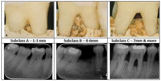
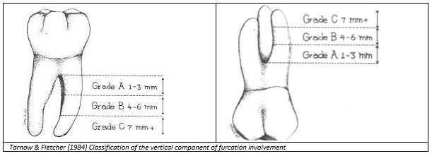
2. Classification of the vertical component of furcation involvement
A sub-classification referring to the vertical bone loss from the furcation fornix was introduced by Tarnow and Fletcher (1984) to complement the horizontal classification (I–III):
Subclass A: vertical bone loss 3 mm or less
Subclass B: vertical bone loss from 4 to 6 mm
Subclass C: bone loss from the fornix of 7 mm or more
Step I: Identify fornix of the furcation on PA radiogram
Step II: Divide root length into thirds (measured from the fornix) – cervical, middle and apical
Step III: Identify the level of bone in the furcation
Step IV: Assess the location of the bone loss in the furcation with respect to the root thirds